Monday, March 23, 2009

What is Tennis Elbow ?


[Extracted from WiseGeek.com]

Tennis elbow, or lateral epicondylitis, is a painful condition that affects the outside or lateral side of the elbow and can cause pain even when gripping or carrying a very light object like a coffee cup. About half of all tennis players will experience tennis elbow at some point in their lives, yet tennis players only account for about 5% of all those who suffer from tennis elbow, as the condition can be brought on by many activities.

Tennis elbow is often confused with two other painful conditions: golfer's elbow or medial epicondylitis, and bursitis. Golfer's elbow is brought about by the same types of activities as tennis elbow, but the pain is primarily felt on the inside of the elbow rather than the outside, while the pain of bursitis is at the back of the elbow.

Tennis elbow will typically cause recurring pain at the outside top of the forearm just below the elbow, and pain may radiate down the arm. Bending the arm, lifting or straightening the arm will also cause pain due to inflamed muscles and tendons. Typical bouts of tennis elbow last 6-12 weeks, but it can also last shorter or longer periods of time.

Tennis elbow is caused by small tears in the tendons and muscles. Tendons anchor muscles to bone. When tendons tear it takes much longer to heal than muscle tissue because tendons receive less blood and oxygen than muscles.

Once tendons and muscles are injured, the site is always vulnerable to re-tearing. Further injury can lead to hemorrhaging and calcium deposits in the tissues. A protein called collagen can also create inflammation that can press on the radial nerve that controls muscles in the arm and hand.

Because tennis elbow is associated with injury to the tendon it is often referred to as tendonitis, however, if the bone itself feels sore and muscles are involved it is epicondylitis or tennis elbow. Although tennis elbow involves inflammation of the tissues at the injury site, it does not cause swelling of the arm, which could be indicative of arthritis, gout or even infection.

The recommended treatment for tennis elbow is to stop whatever activities have caused it and allow the arm to rest until all pain has ceased. At that point massage and exercise may be of value to strengthen the arm against further injury. Slow gentle stretching of the arm for 10-15 minutes before engaging in any activity is also recommended.

For special cases of tennis elbow your doctor may suggest anti-inflammatory creams, injections or even surgery. However surgery is a last resort suggested to less than 3% of those who suffer from tennis elbow. In this case tears in the tendon may be repaired or the tendon may be cut free of the bone to release the stress, though this is rarely chosen as it leaves the tendon useless.


Tennis Elbow (lateral epicondylitis)
Outside of Elbow - Cause & Symptoms

The onset of pain, on the outside (lateral) of the elbow, is usually gradual with tenderness felt on or below the joint's bony prominence. Movements such as gripping, lifting and carrying tend to be troublesome.


Golfer’s Elbow (medial epicondylitis)
Inside of Elbow - Cause & Symptoms

The causes of golfers elbow are similar to tennis elbow but pain and tenderness are felt on the inside (medial) of the elbow, on or around the joint's bony prominence.


Bursitis
Back of Elbow - Cause & Symptoms

Often due to excessive leaning on the joint or a direct blow or fall onto the tip of the elbow. A lump can often be seen and the elbow is painful at the back of the joint.

To prevent tennis elbow from recurring, experts suggest modifying the activity that caused it, stretching or warming up the arm, exercising the arm to build muscle strength, and resting the arm between activities.

If you are experiencing persistent pain in your elbow or arm it is always best to see your doctor in order to have the problem professionally diagnosed and treated

Saturday, March 21, 2009

What is meant by a Ruptured Spleen ?


[Extracted from MayoClinic.Com]

A ruptured spleen is a serious condition that can occur when your spleen experiences a trauma. Without emergency treatment, a ruptured spleen can cause life-threatening bleeding.

Your spleen is located just under your rib cage on your left side, a prime spot for injury. With enough force, a blow to your abdomen — during a sporting mishap, a fist fight or a car accident, for example — might lead to a ruptured spleen.

In the past, ruptured spleens usually were removed in emergency surgery. These days, however, it's common for people with ruptured spleens to be treated instead with several days of hospital care to help the spleen heal.

The spleen helps your body fight infection and filter unwanted material, such as old or damaged blood cells. The spleen also produces red blood cells and certain types of white blood cells. A ruptured spleen may pour a large amount of blood into your abdominal cavity.

Abdominal pain and tenderness are the classic symptoms of a ruptured spleen. If internal bleeding causes your blood pressure to drop, you may become lightheaded or confused. Blurred vision and fainting are possible as well.


When to see a doctor ?

A ruptured spleen is a medical emergency. If you have pain in the left upper abdomen or signs and symptoms of low blood pressure — such as lightheadedness, confusion, blurred vision or fainting — following an injury, seek emergency medical care.
A ruptured spleen is typically caused by a blow to the left upper abdomen or the left lower chest. Sporting mishaps, fist fights and car accidents are common triggers. An injured spleen may rupture soon after the abdominal trauma or, in some cases, days or even weeks after the injury.

An enlarged spleen — which may be caused by various underlying problems, from mononucleosis and other infections to liver disease and blood cancers — may sometimes rupture with trauma to the abdomen. In fact, a ruptured spleen is the most significant complication of mononucleosis.

A ruptured spleen can cause life-threatening bleeding into your abdominal cavity.

In a critical situation, a physical exam may be the only diagnostic test before emergency surgery. In other cases, a sample of fluid from your abdomen may be tested for the presence of blood. If time allows, your doctor may use CT scans or other imaging tests to identify or rule out other possible causes of your symptoms.

Many small and many moderate-sized injuries to the spleen can heal without surgery. You're likely to stay in the hospital for up to a week while doctors observe your condition and provide nonsurgical care, such as blood transfusions, if necessary. If your injury is severe, your spleen may need to be surgically repaired or, in some cases, removed. Surgery to remove your spleen is called splenectomy.

Many splenectomies are done with a small incision and a slender tube equipped with a camera lens and light (laparoscope), but emergency surgery may require an open procedure. If you've lost a large amount of blood, you may need transfusions to improve your circulation.

Life without a spleen

You can live an active life without a spleen, but you're more likely to contract serious or even life-threatening infections. If your spleen is removed, your doctor may recommend a pneumonia vaccine, as well as yearly flu vaccines. In some cases, preventive antibiotics may be recommended as well — especially if you have any other conditions that increase your risk of serious infections.

After a splenectomy, notify your doctor at the first sign of an infection. Make sure anyone caring for you knows that you've had your spleen removed.

What is a Herniated Disc ?


[Extracted from ABOUT.COM]

Many patients with back pain, leg pain, or weakness of the lower extremity muscles are diagnosed with a herniated disc. When a disc herniation occurs, the cushion that sits between the spinal vertebra is pushed outside its normal position. A herniated disc would not be a problem if it weren't for the spinal nerves that are very close to the edge of these spinal discs.

What is the spinal disc ?

The spinal disc is a soft cushion that sits between each vertabrae of the spine. This spinal disc becomes more rigid with age. In a young individual, the disc is soft and elastic, but like so many other structures in the body, the disc gradually looses its elasticity and is more vulnerable to injury. In fact, even in individuals as young as 30, MRIs show evidence of disc deterioration in about 30% of people.

What happens with a 'herniated disc' ?

As the spinal disc becomes less elastic, it can rupture. When the disc ruptures, a portion of the spinal disc pushes outside its normal boundary--this is called a herniated disc. When a herniated disc bulges out from between the vertebrae, the spinal nerves and spinal cord can become pinched. There is normally a little extra space around the spinal cord and spinal nerves, but if enough of the herniated disc is pushed out of place, then these structures may be compressed.


What causes symptoms of a herniated disc ?

When the herniated disc ruptures and pushes out, the nerves may become pinched. A herniated disc may occur suddenly in an event such as a fall or an accident, or may occur gradually with repetitive straining of the spine. Often people who experience a herniated disc already have spinal stenosis, a problem that causes narrowing of the space around the spinal cord and spinal nerves. When a herniated disc occurs, the space for the nerves is further diminished, and irritation of the nerve results.

What are the symptoms of a herniated disc ?

When the spinal cord or spinal nerves become compressed, they don't work properly. This means that abnormal signals may get passed from the compressed nerves, or signals may not get passed at all. Common symptoms of a herniated disc include :


a) Electric Shock Pain
Pressure on the nerve can cause abnormal sensations, commonly experienced as electric shock pains. When the compression occurs in the cervical (neck) region, the shocks go down your arms, when the compression is in the lumbar (low back) region, the shocks go down your legs.

b) Tingling & Numbness
Patients often have abnormal sensations such as tingling, numbness, or pins and needles. These symptoms may be experienced in the same region as painful electric shock sensations.

c) Muscle Weakness
Because of the nerve irritation, signals from the brain may be interrupted causing muscle weakness. Nerve irritation can also be tested by examining reflexes.

d) Bowel or Bladder Problems
These symptoms are important because it may be a sign of cauda equina syndrome, a possible condition resulting from a herniated disc. This is a medical emergency, and your should see your doctor immediately if you have problems urinating, having bowel movements, or if you have numbness around your genitals.

All of these symptoms are due to the irritation of the nerve from the herniated disc. By interfering with the pathway by which signals are sent from your brain out to your extremities and back to the brain, all of these symptoms can be caused by a herniated disc pressing against the nerves.

How is the diagnosis of a herniated disc made ?

Most often, your physician can make the diagnosis of a herniated disc by physical examination. By testing sensation, muscle strength, and reflexes, your physician can often establish the diagnosis of a herniated disc.

An MRI is commonly used to aid in making the diagnosis of a herniated disc. It is very important that patients understand that the MRI is only useful when used in conjunction with examination findings. It is normal for a MRI of the lumbar spine to have abnormalities, especially as people age. Patients in their 20s may begin to have signs of disc wear, and this type of wear would be expected on MRIs of patients in their 40s and 50s. This is the reason that your physician may not be concerned with some MRI findings noted by the radiologist.

Making the diagnosis of a herniated disc, and coming up with a treatment plan depends on the symptoms experienced by the patient, the physical examination findings, and the x-ray and MRI results. Only once this information is put together can a reasonable treatment plan be considered.

Treatment of a herniated disc depends on a number of factors including :

* Symptoms experienced by the patient
* Age of the patient
* Activity level of the patient
* Presence of worsening symptoms

Most often, treatments of a herniated disc begin conservatively, and become more aggressive if the symptoms persist. After diagnosing a herniated disc, treatment usually begins with :

1) Rest & Activity Modification
The first treatment is to rest and avoid activities that aggravate your symptoms. Many disc herniations will resolve is given time. In these cases, it is important to avoid activities that aggravate your symptoms.

2) Ice & Heat Applications
Ice and heat application can be extremely helpful in relieving the painful symptoms of a disc herniation. By helping to relax the muscles of the back, ice and heat applications can relieve muscle spasm and provide significant pain relief.

3) Physical Therapy
Physical therapy and lumbar stabilization exercises do not directly affect the herniated disc, but they can stabilize the lumbar spine muscles. This has an effect of decreasing the load experienced by the disc and vertebrae. Stronger, well balanced muscles help control the lumbar spine and minimize the risk or injury to the nerves and the disc.

4) Anti-Inflammatory Medications
Nonsteroidal anti-inflammatory medications (NSAIDs) are commonly prescribed, and often help relieve the pain associated with a disc herniation. By reducing inflammation, these medications can relieve some pressure on the compressed nerves. NSAIDs should be used under your doctor's supervision.

5) Oral Steroid Medications
Oral steroid medications can be very helpful in episodes of an acute (sudden) disc herniation. Medications used include Prednisone and Medrol. Like NSAIDs, these powerful anti-inflammatory medications reduce inflammation around the compressed nerves, thereby relieving symptoms.

6) Other Medications
Other medications often used include narcotic pain medications and muscle relaxers. Narcotic pain medications are useful for severe, short-term pain management. Unfortunately, these medication can make you drowsy and can be addictive. It is important to use these for only brief periods of time. Muscle relaxers are used to treat spasm of spinal muscles often seen with disc herniations. Often the muscle spasm is worse than the pain from the disc pressing on the nerves.

7) Epidural Steroid Injections
Injections of cortisone can be administered directly in the area of nerve compression. Like oral anti-inflammatory medications, the idea is to relieve the compression on the nerves. When the injection is used, the medication is delivered to the area of the disc herniation, rather than being taken orally and travelling throughout your body.

Is surgery necessary in the treatment of a disc herniation ?
As mentioned, treatment of a disc herniation usually begins with the steps listed above. However, surgical treatment of a herniated disc may be recommended soon after the injury if there is a significant neurological deficit to your problem. Symptoms on pain and sensory abnormalities usually do not require immediate intervention, but patients who have significant weakness, any evidence of cauda equina syndrome, or a rapidly progressing problem may require more prompt surgical treatment.

Most often surgery is recommended if more conservative measures do not relieve your symptoms. Surgery is performed to remove the herniated disc, and free up space around the compressed nerve. Depending on the size and location of the herniated disc, and associated problems (such as spinal stenosis, arthritis, etc.), the surgery can be done by several techniques. In very straightforward cases, endoscopic or microscopic excision of the herniated disc may be possible. However, this is not always recommended, and in some cases, a more significant surgery may need to be performed.

Lumbar Discectomy

A discectomy is a surgery done to remove a herniated disc from the spinal canal. When a disc herniation occurs, a fragment of the normal spinal disc is dislodged. This fragment may press against the spinal cord or the nerves that surround the spinal cord. This pressure causes the symptoms that are characteristic of herniated discs.

The surgical treatment of a herniated disc is to remove the fragment of spinal disc that is causing the pressure on the nerve. This procedure is called a discectomy. The traditional surgery is called an open discectomy. An open discectomy is a procedure where the surgeon uses a small incision and looks at the actual herniated disc in order to remove the disc and relieve the pressure on the nerve.

How is a discectomy performed ?

A discectomy is performed under general anesthesia. The procedure takes about an hour, depending on the extent of the disc herniation, the size of the patient, and other factors. A discectomy is done with the patient lying face down, and the back pointing upwards.

In order to remove the fragment of herniated disc, your surgeon will make an incision over the center of your back. The incision is usually about 3 centimeters in length. Your surgeon then carefully dissects the muscles away from the bone of your spine. Using special instruments, your surgeon removes a small amount of bone and ligament from the back of the spine. This part of the procedure is called a laminotomy.

Once this bone and ligament is removed, your surgeon can see, and protect, the spinal nerves. Once the disc herniation is found, the herniated disc fragment is removed. Depending on the appearance and the condition of the remaining disc, more disc fragments may be removed in hopes of avoiding another fragment of disc from herniating in the future. Once the disc has been cleaned out from the area around the nerves, the incision is closed and a bandage is applied.

What is the recovery from a discectomy ?

Patients often awaken from surgery with complete resolution of their leg pain; however, it is not unusual for these symptoms to take several weeks to slowly dissipate. Pain around the incision is common, but usually well controlled with oral pain medications. Patients often spend one night in the hospital, but are usually then discharged the following day. A lumbar corset brace may help with some symptoms of pain, but is not necessary in all cases.

Gentle activities are encouraged after surgery, such as sitting upright and walking. Patients must avoid lifting heavy objects, and should try not to bend or twist the back excessively. Patients should avoid strenuous activity or exercise until cleared by their doctor.

What are the potential complications of a discectomy ?

The most common problem of a discectomy is that there is a chance that another fragment of disc will herniate and cause similar symptoms down the road. This is a so-called recurrent disc herniation, and the risk of this occurring is about 10-15%.

Most patients find relief of much, if not all, of their symptoms from a discectomy. However, the success of the procedure is about 85-90%, meaning that 10% of patients who undergo a discectomy will still have persistent symptoms. Patients who have symptoms for long periods of time, or severe neurologic deficits (such as significant weakness) are at higher risk of incomplete recovery.

Other risks of surgery include spinal fluid leaks, bleeding, and infection. All of these can usually be treated, but may require a longer hospitalization or additional surgery.

What is endoscopic microdiscectomy ?

Newer techniques may allow your surgeon to perform a procedure called an endoscopic discectomy. In an endoscopic discectomy your surgeon uses special instruments and a camera to remove the herniated disc through very small incisions.

The endoscopic microdiscectomy is a procedure that accomplishes the same goal as a traditional open discectomy, removing the herniated disc, but uses a smaller incision. Instead of actually looking at the herniated disc fragment and removing it, your surgeon uses a small camera to find the fragment and special instruments to remove it. The procedure may not require general anesthesia, and is done through a smaller incision with less tissue dissection. Your surgeon uses x-ray and the camera to "see" where the disc herniation is, and special instruments to remove the fragment.

Endoscopic microdiscectomy is appropriate in some specific situations, but not in all. Many patients are better served with a traditional open discectomy. While the idea of a faster recovery is nice, it is more important that the surgery is properly performed. Therefore, if open discectomy is more appropriate in your situation, then the endoscopic procedure should not be done. Discuss with your doctor if endoscopic microdiscectomy may be appropriate for you.

Friday, March 20, 2009

Capsicum and Health Benefits !


Capsicum helps in curing many diseases that include lowering blood pressure, reducing cholesterol and warding off strokes and heart attacks, speeding up metabolism, treating colds and fevers, preventing cancer and pain control. Capsaicin is a flavorless, odorless chemical concentrated in the veins of chiles and peppers.

Capsicum contains Vitamins A and C, and beta-carotene. It's low in fat, calories and cholesterol. If you thought your diabetic diet was supposed to be boring, try spicing up your cuisine with capsaicin-based products, like hot sauce or jalapeno peppers.


Listed below are some of the health benefits of Capsicum :

* Enhances Metabolism: Capsicum increases the metabolism by lowering triglycerides, which are stored fats in blood corpuscles.This helps in burning calories.

* Anti Oxidant Property: Capsicum is an important source of vitamin A and vitamin C. These two vitamins are powerful antioxidants that help in fighting free radicals in the body and help in treating heart diseases, osteoarthritis, bronchial asthma, cataract etc. The free radicals are responsible for causing cellular damage, damage to nerves and blood vessels.

* Pain Relief: Scientists believe that the compound capsaicin found in capsicum blocks the transmission of pain from the skin to the spinal cord. This is considered helpful in treating neuralgia, pain associated with herpes zoster and postoperative amputation trauma.

* Cancer Prevention: The compound capsaicin is also believed to treat cancer by preventing carcinogens from binding with DNA. As a regular dietary intake, it has been found through various studies that eating capsicum can provide some protection from cancer.

* Prevents Clotting of Blood: Studies suggest that capsicum has the capacity to prevent clotting of blood. Vitamin C is found to play a major role in dissolving blood clots.

* Fights Food-Borne Bacterial Diseases: In a study it has been found that capsaicin content in capsicum is effective in fighting off food-borne bacterial diseases like Vibrio vulnificus bacteria found in raw shellfish.


Capsicum is a vegetable that comes in an exciting range of colors, like green, red and yellow. It is native to America and in fact, has been cultivated by the people of the tropical Americas since thousands of years. Known by a number of names, like chili pepper, bell pepper and red or green pepper, the vegetable is used as a spice, medicine and obviously, a vegetable. Apart from being great in taste, capsicum also holds a great degree of nutritional value and accords numerous health benefits to people. In the following lines, we have listed the nutrition benefits of eating capsicum.


Nutritional Value of Capsicum

a) Carotenoids (Capsanthin, Capsorubin, Carotene and Lutein)
b) Proteins
c) Fats
d) Vitamin A
e) Vitamin C
f) Capsaicinoid Oleoresin
g) Provitamins E, P, B1, B2 and B3
h) Steroidal Alkaloidal Glycosides (Solanine and Solasadine)
i) Scopoletin (Coumarin)

Health & Nutrition Benefits of Eating Capsicum

1) Beneficial to the mucous membranes, eyes and skin
2) Helps wards off infection
3) Promotes cardiovascular health, by helping lower blood pressure
4) Has antioxidant properties that neutralize the free radicals responsible for damaging tissue and cells
5) Has anti-inflammatory properties
6) Helps reduce cholesterol and thus, is effective in warding off strokes and heart attacks
7) Speeds up the metabolism and helps burn more calories
8) Good for treating colds and fevers
9) Stimulates stomach secretions and improves digestion
10) Lowers triglycerides
11) Has a laxative effect
12) Triggers the release of endorphins
13) Good for diabetics

Sunday, March 15, 2009

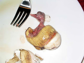
What is Balut ?


Balut is an egg that has been fertilized and contains a partially developed embryo. In countries where the balut is considered to be a delicacy, a duck egg or a chicken egg may be used to prepare the dish. The mode of preparation for balut is very simple. Generally, the egg is boiled and eaten directly from the shell, in much the same manner that boiled and unfertilized eggs are eaten around the world.

In countries where balut is commonly served, the fertilized duck egg or chicken egg is considered to be a superior source of protein. The boiled embryo is also considered to add flavor and texture to the egg. A balut may be enjoyed as a quick snack along with beer or some form of ale, or be employed as the centerpiece for a meal.

Along with the basic method of boiling the fertilized chicken egg or duck egg, it is not uncommon for the exposed contents of the shell to be doused with vinegar, chili peppers, or a variety of spices. Some prefer to not eat the white sections remaining in the shell, while others consume all the contents found within the egg proper. Balut can also be included as a center for baked pastries, as well as mixed into an egg batter and fried into an entrée.

Balut is developed by allowing the fertilized egg to begin developing the embryo for several days. During this period, the egg is kept warm. Generally, eight or nine days of consistent warmth are sufficient to allow the embryo to develop. At this juncture, the balut is ready for boiling. After boiling the balut, the contents of the egg can be used in a number of different recipes. Generally, the balut is always served while still warm.

---------------------------------------------------------------------


Fertilized duck eggs sold as balut in the U.S. range from 16 to 20 days in age. The older the egg, the larger the chick and the more pronounced its feathers, bones, and beak. An embryo at 17 days has beak and feathers which are more developed at 20 days. Normally, after being fertilized, a chick hatches after 26 to 28 days of incubation.

The taste of the egg also depends on the breed of the laying duck. Different breeds of ducks supposedly produce balut varying in taste, with Muscovy ducks being hailed by some as the "cream of the crop". The kind of balut sold in the U.S. is made from duck eggs. Chicken eggs may be made into balut as well, but duck eggs are preferred by the majority of Filipino Americans since they are larger and thought to be better in taste.


But whether the fertilized egg is chicken or duck, there are two types of balut. One is called mamatong by Filipinos. Mamatong balut has the embryo floating on top of the white and yolk and the consumer can easily detect it. Roughly translated to mean "the float," mamatong occurs between 14 to 16 days. The second is balut sa puti where the embryo is wrapped by a thin, whitish membrane and one cannot tell whether there is an embryo or not. In balut sa puti, the embryo is hidden by the albumen's white film. Balut sa puti is 17 to 18 days old and it is the preferred favorite of Filipinos in the U.S. and in the Philippines. A folk belief in the Philippines lets people know if an egg has developed into mamatong or balut sa puti. One takes a balut egg and drops it in water. If it floats, it is mamatong, but if it sinks, it is balut sa puti.

Just how good and fresh a balut is after it has been boiled can be determined by its broth, called "soup" by balut eaters. After cracking a hole in the wide part of the shell, the consumer usually sips the broth before he or she eats any part of the tiny chick and remaining yolk. If the balut is good, its soup has a sweet, clean taste. Fresh balut can be good for ten days to two weeks. Cooked balut if stored in the refrigerator will last for as long as a month. But the longer the balut is in the refrigerator, the more likely that its liquid will be dried out.

Saturday, March 7, 2009

What is Heavy Metal Poisoning ?

[Extracted from The Free Dictionary by Farlex]

Definition
Heavy metal poisoning is the toxic accumulation of heavy metals in the soft tissues of the body.

Description
Heavy metals are chemical elements that have a specific gravity (a measure of density) at least five times that of water. The heavy metals most often implicated in accidental human poisoning are lead, mercury, arsenic, and cadmium. More recently, thallium has gained some attention in the media as the poison used in several murder cases in the 1990s. Some heavy metals, such as zinc, copper, chromium, iron, and manganese, are required by the body in small amounts, but these same elements can be toxic in larger quantities.

Heavy metals may enter the body in food, water, or air, or by absorption through the skin. Once in the body, they compete with and displace essential minerals such as zinc, copper, magnesium, and calcium, and interfere with organ system function. People may come in contact with heavy metals in industrial work, pharmaceutical manufacturing, and agriculture. Children may be poisoned as a result of playing in contaminated soil. Lead poisoning in adults has been traced to the use of lead-based glazes on pottery vessels intended for use with food, and contamination of Ayurvedic and other imported herbal remedies. Arsenic and thallium have been mixed with food or beverages to attempt suicide or poison others.

Another form of mercury poisoning that is seen more and more frequently in the United States is self-injected mercury under the skin. Some boxers inject themselves with mercury in the belief that it adds muscle bulk. Metallic mercury is also used in folk medicine or religious rituals in various cultures. These practices increase the risk of mercury poisoning of children in these ethnic groups or subcultures.

Causes and symptoms
Symptoms will vary, depending on the nature and the quantity of the heavy metal ingested. Patients may complain of nausea, vomiting, diarrhea, stomach pain, headache, sweating, and a metallic taste in the mouth. Depending on the metal, there may be blue-black lines in the gum tissues. In severe cases, patients exhibit obvious impairment of cognitive, motor, and language skills. The expression "mad as a hatter" comes from the mercury poisoning prevalent in 17th-century France among hatmakers who soaked animal hides in a solution of mercuric nitrate to soften the hair.

Diagnosis
Heavy metal poisoning may be detected using blood and urine tests, hair and tissue analysis, or x ray. The diagnosis is often overlooked, however, because many of the early symptoms of heavy metal poisoning are nonspecific. The doctor should take a thorough patient history with particular emphasis on the patient's occupation.
In childhood, blood lead levels above 80 ug/dL generally indicate lead poisoning, however, significantly lower levels (>30 ug/dL) can cause mental retardation and other cognitive and behavioral problems in affected children. The Centers for Disease Control and Prevention considers a blood lead level of 10 ug/dL or higher in children a cause for concern. In adults, symptoms of lead poisoning are usually seen when blood lead levels exceed 80 ug/dL for a number of weeks.

Blood levels of mercury should not exceed 3.6 ug/dL, while urine levels should not exceed 15 ug/dL. Symptoms of mercury poisoning may be seen when mercury levels exceed 20 ug/dL in blood and 60 ug/dL in urine. Mercury levels in hair may be used to gauge the severity of chronic mercury exposure.

Since arsenic is rapidly cleared from the blood, blood arsenic levels may not be very useful in diagnosis. Arsenic in the urine (measured in a 24-hour collection following 48 hours without eating seafood) may exceed 50 ug/dL in people with arsenic poisoning. If acute arsenic or thallium poisoning is suspected, an x ray may reveal these substances in the abdomen (since both metals are opaque to x rays). Arsenic may also be detected in the hair and nails for months following exposure.
Cadmium toxicity is generally indicated when urine levels exceed 10 ug/dL of creatinine and blood levels exceed 5 ug/dL.

Thallium poisoning often causes hair loss (alopecia), numbness, and a burning sensation in the skin as well as nausea, vomiting, and dizziness. As little as 15-20 mg of thallium per kilogram of body weight is fatal in humans; however, smaller amounts can cause severe damage to the nervous system.

Treatment
When heavy metal poisoning is suspected, it is important to begin treatment as soon as possible to minimize long-term damage to the patient's nervous system and digestive tract. Heavy metal poisoning is considered a medical emergency, and the patient should be taken to a hospital emergency room.

The treatment for most heavy metal poisoning is chelation therapy. A chelating agent specific to the metal involved is given either orally, intramuscularly, or intravenously. The three most common chelating agents are calcium disodium edetate, dimercaprol (BAL), and penicillamine. The chelating agent encircles and binds to the metal in the body's tissues, forming a complex; that complex is then released from the tissue to travel in the bloodstream. The complex is filtered out of the blood by the kidneys and excreted in the urine. This process may be lengthy and painful, and typically requires hospitalization. Chelation therapy is effective in treating lead, mercury, and arsenic poisoning, but is not useful in treating cadmium poisoning. To date, no treatment has been proven effective for cadmium poisoning. Thallium poisoning is treated with a combination of Prussian blue (potassium ferric hexacyanoferrate) and a diuretic, because about 35% of it is excreted in the urine; however, if treatment is not started within 72 hours of ingesting the poisoning, damage to the patient's nervous system may be permanent.

In cases of acute mercury, arsenic, or thallium ingestion, vomiting may be induced. Activated charcoal may be given in cases of thallium poisoning. Washing out the stomach (gastric lavage) may also be useful. The patient may also require treatment such as intravenous fluids for such complications of poisoning as shock, anemia, and kidney failure.

Patients who have taken arsenic, thallium, or mercury in a suicide attempt will be seen by a psychiatrist as part of emergency treatment.

Prognosis
The chelation process can only halt further effects of the poisoning; it cannot reverse neurological damage already sustained.

Prevention
Because arsenic and thallium were commonly used in rat and insect poisons at one time, many countries have tried to lower the rate of accidental poisonings by banning the use of heavy metals in pest control products. Thallium was banned in the United States as a rodent poison in 1984. As a result, almost all recent cases of arsenic and thallium poisoning in the United States were deliberate rather than accidental.

Because exposure to heavy metals is often an occupational hazard, protective clothing and respirators should be provided and worn on the job. Protective clothing should then be left at the work site and not worn home, where it could carry toxic dust to family members. Industries are urged to reduce or replace the heavy metals in their processes wherever possible. Exposure to environmental sources of lead, including lead-based paints, plumbing fixtures, vehicle exhaust, and contaminated soil, should be reduced or eliminated.

Friday, March 6, 2009

What is Anal Itching ?


Anal itching is itching around your anus — the canal that's the outlet for your rectum. The itch, located in your anus or on the skin just around your anus, is often intense and may be accompanied by a strong urge to scratch. You may find anal itching to be an embarrassing and uncomfortable situation.

Also called pruritus ani, anal itching has many causes. Numerous factors may cause anal itching to be more intense — including moisture, abrasion caused by your clothing and the pressure of sitting.

Anal itching is a common problem that many people have experienced. Don't be afraid to talk with your doctor about this condition. With proper treatment and self-care measures, most people can achieve complete relief from anal itching.

Symptoms
Anal itching is associated with other similar symptoms in and around the anus, including :

* Burning
* Soreness or pain

The itching and irritation in and around your anus may be a temporary condition, or it can be a more persistent and bothersome problem. For some people, the irritation is so intense that the urge to scratch is both irresistible and embarrassing.

Causes
Most cases of anal itching are caused by a harmless problem. Occasionally, however, anal itching can be a sign of more-serious medical issues. Possible causes of anal itching include :

** Dry skin. As you age, skin in and around your anus is more prone to dryness. Dry skin can cause persistent, intense anal itching.

** Too much moisture. Moisture around your anus from excessive sweating or from moist, sticky stools can be irritating. Anal itching can also be caused by frequent diarrhea or the escape of small amounts of stool (fecal incontinence).

** Excessive washing. Excessive wiping with dry, harsh toilet paper or excessive scrubbing with harsh soaps can cause or aggravate anal itching. Failure to rinse away the soap completely also may cause irritation.

** Chemical irritants. Certain laundry soaps, colognes, douches and birth control products contain chemicals that can irritate skin in and around your anus. Scented or colored toilet paper can be irritating to people with sensitive skin.

** Food irritants. Anal itching may be the result of irritating chemicals in some foods, such as those found in spices and hot sauces. Similarly, some foods may directly or indirectly irritate your anus as they exit your colon. Common culprits include chocolate, alcohol, tomatoes, nuts and popcorn. Consuming certain beverages, including milk or caffeinated drinks, may cause some people to experience diarrhea followed by anal itching.

** Medications. Anal itching may be a side effect of certain medications, including some antibiotics, that can cause frequent diarrhea.

** Overuse of laxatives. Excessive or improper use of laxatives can lead to chronic diarrhea and the risk of anal irritation and itching.

** Hemorrhoids. Hemorrhoids are engorged veins located just under the membrane that lines the lowest part of your rectum and anus. They often occur as a result of straining during a bowel movement. Anal itching can be a symptom of hemorrhoids.

** Infections. Sexually transmitted diseases may also involve the anus and can cause anal itching. In children, the parasite that causes pinworms can cause persistent anal itching. Other parasites may cause similar itching.

** Skin disorders. Common skin problems — such as psoriasis, seborrhea and eczema — can involve and irritate the area in and around your anus.

** Yeast infections. This common infection, which usually affects women, can irritate your genital and anal areas.

** Anal abrasions and fissures. An anal abrasion is a small tear in your anus, usually caused by forced bowel movements through a tight anus. An anal fissure is a deeper tear. Both conditions can cause anal itching, as well as painful bowel movements and bleeding.

** Anal tumors. Rarely, benign or cancerous tumors in or around the anus may be a cause of anal itching.

** Other causes. Anal itching may be related to anxiety or stress. Sometimes, the cause remains undetermined.

Contributing to the problem
Although anal itching is almost never a problem of cleanliness, your own actions may contribute to the problem. Whatever the cause of anal itching, your natural reaction is to scratch the area. But scratching worsens the problem by removing superficial layers of skin. In addition, the natural tendency in response to an irritation is to wash the area frequently with soap and a washcloth. However, excessive washing can aggravate the condition by removing your skin's natural protective oils.

Most anal itching doesn't require medical care. However, see your doctor if :

* Anal itching is severe or prolonged
* There's bleeding from the rectum
* The itch can't be easily explained

Persistent anal itching may be related to a skin condition or other health problem that requires medical treatment.

What is Cholera ?

[Extracted from WiseGeek.com]

Cholera is a serious infectious disease caused by the bacteria Vibrio cholerae, which affects the intestinal system of the body. An infected person experiences severe vomiting, explosive diarrhea and severe dehydration. Without immediate medical treatment, cholera may result in death within four to twelve hours after symptoms begin. Due to a large loss of body fluids, cholera is gruesome in the way that it leaves survivors in their physical appearance, as well as in the biological toll it takes on the body.

Cholera is very contagious. It is spread by the unintentional consumption of infected feces that contaminate food and water. It can also be spread through human to human contact. Some people have been infected with cholera by eating raw or undercooked shellfish.

Cholera is easily treated with fluids and antibiotics. When antibiotics are unavailable, which is commonly the case in areas that are plagued by cholera, a simple mixture of water and glucose for rehydration is life saving. Cholera usually resolves itself after a period of time; the danger is the severe dehydration that quickly causes death.

Similar to the bubonic plague which has showed its face time and again, cholera is a plague that is suspected to be an ancient disease. It has unquestionably reoccurred in seven pandemics since 1817, including in Europe and the United States. The most recent pandemic began in Asia in 1961 and continues to the present day in Africa.

In developing countries, cholera is prevalent in areas that do not enjoy sanitary living conditions because of poverty and a lack of resources. In pandemic regions, even sanitary conditions may not prevent further outbreak. Many people do not receive information on how the disease is spread, fear seeking medical help or simply do not have access to any kind of treatment.

Cholera is not common in developed countries due to the availability of medical treatment, regulated heath standards, clean water and effective sewage systems. Cases of cholera in developed countries are the result of raw shellfish or people who have contracted the illness while traveling. Those traveling to countries that have cholera epidemics should take careful precautions to prevent infection.

Precautions for travelers include boiling water before drinking or washing. Water can also be treated with chlorine or iodine. Frequent hand washing with clean water, especially after using the toilet, is imperative. Travelers are also advised to ensure their food is fully cooked and hot, peeling their own fruit and avoiding raw vegetables, including salads. Raw and undercooked shellfish should also be avoided.

Cholera is a disease that causes social stigmas and stereotypes in epidemic areas. Indigenous peoples who commonly fall victim to cholera fear the status of social outcast more than the disease itself. Throughout history, communities that have had outbreaks of cholera are marked as diseased and dirty even after they are no longer at risk for spreading the disease.

What are Antibiotics ?

[Extracted from WiseGeek.com]

Antibiotics are medicinal products that have an anti-bacterial effect - they either kill bacteria in the system or keep them from reproducing, allowing the infected body to heal by producing its own defenses and overcome the infection. When antibiotics were isolated in the mid-twentieth century, they were widely hailed as 'wonder drugs' and indeed, formerly life-threatening infections could now be easily cured within a few days.

The most widely known antibiotic is perhaps penicillin, famously made from mold. When it was introduced, many of the sexually transmitted diseases such as gonorrhea went from being a shameful and life changing event to an embarrassing trip to the doctors.

One of the most prevalent and unstoppable myths about antibiotics is that they can cure a cold. Antibiotics work against bacterial infections, and colds are caused by viruses - therefore, a course of antibiotics will do nothing but perhaps kill off the body's own population of beneficial bacteria, leaving the cold to run its natural course. Nevertheless, patients often pressure their doctors into prescribing antibiotics when they come down with a cold or the flu.

One side effect of taking antibiotics for an infection is that it can leave the body defenseless against other non-bacterial types of infections, and for many women, this means a yeast infection, which is fungal. The fungus responsible for yeast infections is always present, and is kept from spreading by helpful bacteria in the digestive tract, which the antibiotics kill, or at least curtail, leaving the fungus to spread. Over-the-counter yeast infection products will usually work to alleviate a yeast infection.

Widespread use of antibiotics for non-medicinal purposes, such as in cattle feed and in antibacterial hand soaps, is causing concern in the medical and pharmaceutical industries, since it is responsible for the evolution of antibiotic resistant bacteria. Washing your hands with an antibacterial soap may make you feel cleaner, but any bacteria that survives the scrub is part of the bacterial population that is immune to that particular antibiotic, and thus all its descendants will be too. This has led to an "arms race" between bacteria and the drug manufacturers, with some antibiotics being held back or only minimally prescribed to prevent bacteria in the "wild" from adapting to them.

Antibiotics may seem like a recent innovation of the past hundred years, but indeed, many ancient civilizations had some understanding of the principle of antibiotics, and many herbs have anti-bacterial effects. One of the most widely known is common garlic. It is so effective at countering bacteria that over-consumption of garlic can have the same effect as antibiotics at leaving the body prey to non-bacterial infections by reducing the body's "good bacteria" population.

What is Vibrio Bacteria ?

[Extracted from RevolutionHealth.com]


What is Vibrio vulnificus food poisoning ?

Vibrio vulnificus food poisoning is caused by Vibrio vulnificus, a bacterium that lives in warm seawater. The condition is rare.

What causes Vibrio vulnificus food poisoning ?
Vibrio vulnificus food poisoning occurs when you eat seafood infected with the bacteria or you have an open wound that is exposed to them. The bacteria are frequently found in oysters and other shellfish in warm coastal waters during the summer months. People with weak immune systems, especially those with long-term (chronic) liver disease, are at greater risk for developing the condition than others.


What are the symptoms ?
In healthy people, Vibrio vulnificus food poisoning can cause vomiting, diarrhea, and abdominal pain. In people who have weak immune systems, the bacteria can infect the bloodstream, causing a severe and life-threatening illness. Symptoms include fever and chills, decreased blood pressure (septic shock), and blistering skin wounds. The infection is especially dangerous to people with long-term (chronic) liver disease.

If an open wound is exposed to the bacteria (such as from warm seawater), it may result in the skin breaking open and sores developing. People with weak immune systems are at risk for the bacteria moving into the bloodstream.

How is Vibrio vulnificus food poisoning diagnosed ?
Vibrio vulnificus food poisoning is diagnosed based on a medical history and a physical exam. Your health professional will ask you questions about your symptoms, foods you have recently eaten, and your work and home environments. If you have eaten raw seafood, especially oysters, your health professional may do a stool, wound, or blood culture.


How is it treated ?
You treat Vibrio vulnificus food poisoning by managing any complications until it passes. Dehydration caused by diarrhea and vomiting is the most common complication. In people with weak immune systems, or in people with severe symptoms, antibiotics may be used.

To prevent dehydration, take frequent sips of a rehydration drink (such as Lytren, Rehydralyte, or Pedialyte). Try to drink a cup of water or rehydration drink for each large, loose stool you have. Sports drinks, soda pop, and fruit juice contain too much sugar and not enough of the important electrolytes that are lost through diarrhea and should only be taken alternately with a rehydration drink.

Try to stick to your normal diet as much as possible. Eating your usual diet will help you to get enough nutrition. Doctors believe that eating a normal diet will also help you feel better faster. But try to avoid foods that are high in fat and sugar. Also avoid spicy foods, alcohol, and coffee for 2 days after all symptoms have disappeared.


How can I prevent Vibrio vulnificus food poisoning ?
The best way to prevent this type of food poisoning is to not eat raw oysters or other raw shellfish and to cook all shellfish (oysters, clams, mussels) thoroughly.

Boil shucked oysters at least 3 minutes, or fry them in oil at least 10 minutes at 375 F. For shellfish in the shell, either :

Boil until the shells open and continue boiling for 5 more minutes, or
Steam until the shells open and then continue cooking for 9 more minutes.
Do not eat those shellfish that do not open during cooking.

You should also :

* Avoid cross-contamination of cooked seafood and other foods with raw seafood and juices from raw seafood. Don't prepare them in the same place; and don't use the same cutting board when preparing them.

* Eat shellfish immediately after cooking, and refrigerate leftovers.

* Avoid exposing open wounds or broken skin to warm saltwater or brackish water or to raw shellfish harvested from such waters.

* Wear protective clothing, such as gloves, when handling raw shellfish.

+++++++++++++++++++++++++++++++++++++++++++++++++++++++++++++


There was a rare incident which happened in Singapore and was reported in 'The Raw Story', in March 2007. Read more at this website ......

http://rawstory.com/news/dpa/Man_dies_after_being_pricked_by_cra_03212007.html

Tuesday, March 3, 2009

What is Shaken Baby Syndrome (SBS) ?


[Extracted from MedicineNet.com]

Shaken baby syndrome is the term that is used to describe a form of child abuse caused by vigorously shaking an infant, often in anger, to get a child to stop crying or whining. It usually occurs in children less than 01 year of age, and the violent shaking often results in severe and permanent brain injury, spinal-cord injuries, bleeding in the eyes (retinal hemorrhages), and even death.

How common is shaken baby syndrome ?

There are no accurate statistics, but experts estimate the incidence to be between 1,000 to 1,500 infants per year. According to the Centers for Disease Control and Prevention, of the almost 2,000 children who die from abuse or neglect each year, shaken baby syndrome accounts for 10%-12% of them. Most commonly, the victim of shaken baby syndrome is between 03 and 08 months old; however, it has been reported in newborns and in children up to 04 years of age. In addition, 25% of all children diagnosed with shaken baby syndrome die from their injuries.

What causes shaken baby syndrome ?

Infants have very weak neck muscles and large and heavy heads in proportion to their bodies. In addition, because the infant brain is immature and needs room to grow, there is naturally a virtual space between the skull and brain to allow for development. Violently shaking an infant can cause the brain to move within the skull, resulting in cerebral contusions (bruising of brain tissue) and shearing (tearing) of blood vessels. Most commonly, the injuries associated with shaken baby syndrome include bleeding around the brain (subdural and subarachnoid hemorrhages), bleeding in the eyes (retinal hemorrhages), and spinal cord or neck injuries. Often infants will also have evidence of other non-accidental injuries, including unexplained bruises, rib fractures, or extremity fractures.

What are the symptoms and signs of shaken baby syndrome ?

The injuries associated with shaken baby syndrome may not be immediately noticeable. Infants may present with nonspecific complaints, such as irritability or vomiting. These symptoms are caused by the developing increased pressure within the brain (intracranial pressure) caused by brain hemorrhages and swelling. These infants often develop additional symptoms, such as lethargy, breathing difficulties, and seizures.

What are the treatments for shaken baby syndrome ?

Infants with injuries of shaken baby syndrome require emergency care, including respiratory support and surgery. Often these infants require draining of the blood around the brain to decrease the ongoing brain injuries associated with brain swelling. Additional treatments may be required as well, including obtaining ophthalmologic and neurological evaluations.

What is the prognosis for shaken baby syndrome ?

Infants who have suffered injuries as a result of this type of child abuse have a poor prognosis. Of those infants who survive, many will have permanent brain damage, retinal injuries, and blindness as well as and neck and spinal injuries. Unfortunately, the damage to the nervous system is too frequently permanent.

Can shaken baby syndrome be caused accidentally ?

Shaken baby syndrome is almost always a result of child abuse, often perpetrated by a parent or caregiver who shakes an infant angrily in response to persistent crying. In very rare cases, the injuries associated with shaken baby syndrome may be caused by accidental actions, such as jogging with a newborn baby in a backpack. It does not result from gentle play or bouncing a child on a knee. Even in those rare accidental cases, the injuries are rarely as severe as those cases associated with non-accidental trauma from shaken baby syndrome.

Can shaken baby syndrome be prevented ?

The following are guidelines to prevent child abuse and shaken baby syndrome

a) Never shake an infant or child.

b) Avoid holding your infant during arguments.

c) Avoid disciplining your child when you are angry.

d) Report abuse to the local police or to your state's child protective services if you suspect that a child in your home or someone you know is a victim of child abuse.

e) If you find yourself becoming increasingly short-tempered around your infant or child, take a break and ask a friend or family member for help.

f) Support the passage of the shaken baby syndrome Prevention Act (S 1204, HR 2052) introduced last year by writing your state senator and congressman.

Shaken baby syndrome is a form of child abuse in which violent shaking of an infant causes the brain to move inside the skull, causing bruising of the brain and tearing of blood vessels. The shaking results in bleeding around the brain, bleeding in the eyes, spinal cord and neck injuries, bruises, and rib fractures. Infants who survive may suffer brain damage and blindness.